(Untitled)
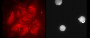
Dr. Belal Tafech (Hedtrich lab, Pharmaceutical Sciences) uses Perkin Elmer Spinning disk to perform live cell imaging of the restricted cilia beating of cystic fibrosis lung models built at the Hedtrich lab (left) and the Brownian motion of 10 um spherical particles in water (Right).
(Untitled)
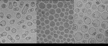
Dr. Arash Momeni (Cullis lab, Biochemistry) performs CryoTEM using the Tecnai G20 200kV TEM to examine the lipid nanoparticles. Left: 5 nm iron oxide nanoparticles and anticancer drug in lipid nanoparticles, Middle: mRNA loaded lipid nanoparticles, Right: 5 nm gold nanoparticles in lipid nanoparticles.

(Untitled)
Sean Ritter, a PhD student in Wasteneys lab (Botany), uses the PerkinElmer Spinning disk to look at cell file organization in the Arabidopsis root labelled with the plasma membrane marker LTI6b:GFP (left) and microtubule organization in the clasp-1 mutant as labelled with pUBQ1:neonGreen-TUB2 (Right).